Shoulder Muscle Anatomy Diagram : FULL VIEW OF BODY MUSCLE CHART SHOULDER MUSCLE CHART ARM ... - Plus, exercises for training them.
byAdmin•
0
Shoulder Muscle Anatomy Diagram : FULL VIEW OF BODY MUSCLE CHART SHOULDER MUSCLE CHART ARM ... - Plus, exercises for training them.. To keep things simple, we can divide the shoulder into layers. The shoulder has about eight muscles that attach to the scapula, humerus, and clavicle. Muscles of the shoulder : The pectoralis major muscle, the deltoid muscle and the muscles of the rotator cuff are some of the muscles that move the arm at the glenohumeral joint. Shoulder muscle anatomy shoulder anatomy this large muscle in the back of the upper arm helps straighten the arm.
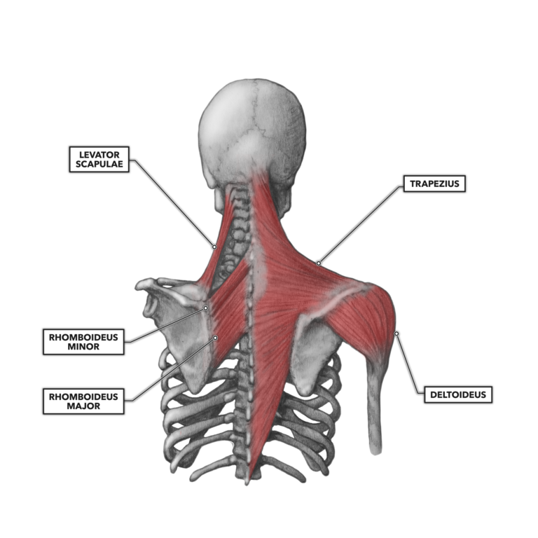
Rhomboid muscles, trapezius muscle and serratus anterior muscle are a few of the scapular stabilizing muscles. There are many nerves and blood vessels that supply the muscles and bones of the shoulder. Shoulder muscles move the shoulder blades and upper arm bones. Deltoids anatomy when most people think of the Anatomyzone 1 free online anatomy resource.
CrossFit | Shoulder Muscles, Part 2: Posterior Musculature from www.crossfit.com Muscles of the shoulder : A numeric illustration was then added to show bone anatomy, muscles attachments, ligaments and muscle layers of the rotator cuff. These muscles form the outer shape of the shoulder and underarm. A muscle contracts to move bones; Anatomy of the shoulder muscles and ligament the rotator cuff and muscles are also shown. The tendons are the attachment of the muscle to the bone. Image via lh4.googleusercontent.com you can see in the shoulder muscle diagrams that the shoulder is one of the largest and most complex joints in the body. Explore a full rotator cuff tear.
It's also responsible for arm abduction, extension, and lateral rotation.
The shoulder is a complex combination of bones and joints where many muscles act to provide the widest range of motion of any part of the body. The right scapula from the front and back side. Ebraheim's educational animated video describes muscle anatomy of the shoulder girdle and anatomy of the shoulder joint.anatomy of the shoulder muscles a. Image via lh4.googleusercontent.com you can see in the shoulder muscle diagrams that the shoulder is one of the largest and most complex joints in the body. Symptoms of rotator cuff tendonitis typically get worse over time. The shoulder muscles consist of the deltoids and the rotator cuff group.the deltoids are the muscles that can be seen on the outside of the body, whilst the rotator cuff group is found within the shoulder joint itself, providing structural support and allowing the shoulder to perform many functions. The muscles of the shoulder bridge the transitions from the torso into the head/neck area and into the upper extremities of the arms and hands. Male shoulder ligaments and biceps muscles isolated in skeleton labeled chart on white labeled human anatomy diagram of male shoulder ligaments, connective tissue and biceps muscles isolated within the skeletal system frontal anterior view on a white background. The tendons are the attachment of the muscle to the bone. Shoulder muscles move the shoulder blades and upper arm bones. Explore a full rotator cuff tear. This article covers the anatomy of the biceps brachii muscle its origins and insertions innervation and functions. A numeric illustration was then added to show bone anatomy, muscles attachments, ligaments and muscle layers of the rotator cuff.
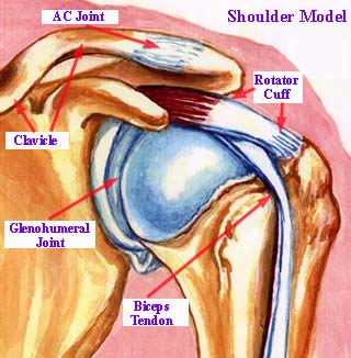
The following is an overview of the shoulder muscle anatomy. Rotator cuff tendonitis is the inflammation or irritation of the tendons and muscles in the shoulder joint. The muscles of the shoulder bridge the transitions from the torso into the head/neck area and into the upper extremities of the arms and hands. For that reason, and because of the dexterity of the shoulder joint itself, the musculature of the shoulder is complex, ranging from massive prime mover muscles to finer stabilizer and fixator muscles. The acromioclavicular joint is formed by an articulation between the lateral end of the clavicle and the acromion process of the scapula.
Male Anatomy Diagram Front View - Male Skeleton Internal ... from media.istockphoto.com Here i have explained the shoulder muscles and in my previous video i have talked about the shoulder j. Bone, then ligaments of the joint capsule, with tendons and muscles on top. The right scapula from the front and back side. To keep things simple, we can divide the shoulder into layers. Anatomyzone 1 free online anatomy resource. The large shoulder muscles are responsible for most of the shoulder's work. To further reinforce the shoulder, the four muscles of the rotator cuff extend from the scapula and surround the head of the humerus to both rotate the arm and prevent dislocation. The shoulder muscles consist of the deltoids and the rotator cuff group.the deltoids are the muscles that can be seen on the outside of the body, whilst the rotator cuff group is found within the shoulder joint itself, providing structural support and allowing the shoulder to perform many functions.
Shoulder flexion is movement of the shoulder in a forward motion.
The large shoulder muscles are responsible for most of the shoulder's work. Numerous muscles help stabilize the three joints of. Muscles of the shoulder : The upper part of the trapezius muscle also helps shrug the shoulder. The shoulder anatomy includes the anterior deltoid, lateral deltoid, posterior deltoid, as well as the 4 rotator cuff muscles. Four of them are found on the anterior aspect of the shoulder, whereas the rest are located on the shoulder's posterior aspect and in the back. The shoulder muscles consist of the deltoids and the rotator cuff group.the deltoids are the muscles that can be seen on the outside of the body, whilst the rotator cuff group is found within the shoulder joint itself, providing structural support and allowing the shoulder to perform many functions. Ebraheim's educational animated video describes muscle anatomy of the shoulder girdle and anatomy of the shoulder joint.anatomy of the shoulder muscles a. This article covers the anatomy of the biceps brachii muscle its origins and insertions innervation and functions. The rotator cuff is a group of four muscles and tendons that surround the glenohumeral joint. Bone, then ligaments of the joint capsule, with tendons and muscles on top. Image via lh4.googleusercontent.com you can see in the shoulder muscle diagrams that the shoulder is one of the largest and most complex joints in the body. The pectoralis major muscle, the deltoid muscle and the muscles of the rotator cuff are some of the muscles that move the arm at the glenohumeral joint.
This, to me, is the best way to show you how much your horse moves as a unit. Plus, exercises for training them. Rhomboid muscles, trapezius muscle and serratus anterior muscle are a few of the scapular stabilizing muscles. Bone, then ligaments of the joint capsule, with tendons and muscles on top. Related posts of shoulder muscles and tendons diagram muscle structure of the knee.
Shoulder Joint Anatomy from www.bone-and-joint-pain.com Subscapularis, supraspinatus, infraspinatus and teres minor. It stabilizes the shoulder and holds the head of the humerus in the glenoid a shallow cavity in the scapula. The following is an overview of the shoulder muscle anatomy. Symptoms of rotator cuff tendonitis typically get worse over time. Image via lh4.googleusercontent.com you can see in the shoulder muscle diagrams that the shoulder is one of the largest and most complex joints in the body. Shoulder flexion is movement of the shoulder in a forward motion. It is rimmed with soft tissue called the labrum that makes a deeper socket that molds to fit the humeral head. So this is my second video about the shoulder anatomy.
This, to me, is the best way to show you how much your horse moves as a unit.
The acromioclavicular joint is formed by an articulation between the lateral end of the clavicle and the acromion process of the scapula. Learn about these muscles, their origin and insertion points, and their functional anatomy. The upper part of the trapezius muscle also helps shrug the shoulder. The shoulder is a complex combination of bones and joints where many muscles act to provide the widest range of motion of any part of the body. Trapezius is responsible for elevating the shoulder blade and rotating it during arm abduction. Here i have explained the shoulder muscles and in my previous video i have talked about the shoulder j. Ebraheim's educational animated video describes muscle anatomy of the shoulder girdle and anatomy of the shoulder joint.anatomy of the shoulder muscles a. Subscapularis, supraspinatus, infraspinatus and teres minor. The shoulder has about eight muscles that attach to the scapula, humerus, and clavicle. The shoulder joint is formed where the humerus (upper arm bone) fits into the scapula (shoulder blade), like a ball and socket. Shoulder muscle anatomy shoulder anatomy this large muscle in the back of the upper arm helps straighten the arm. So this is my second video about the shoulder anatomy. Explore a full rotator cuff tear.
Explore a full rotator cuff tear shoulder anatomy diagram. The shoulder joint is formed where the humerus (upper arm bone) fits into the scapula (shoulder blade), like a ball and socket.